
Neonatal Intestinal Obstruction

99% of healthy full-term infants pass their first stool or meconium within 24 hours of birth and all healthy term neonates should do so by 48 hours.
With preterm infants the length of time can extend up to 9 days.
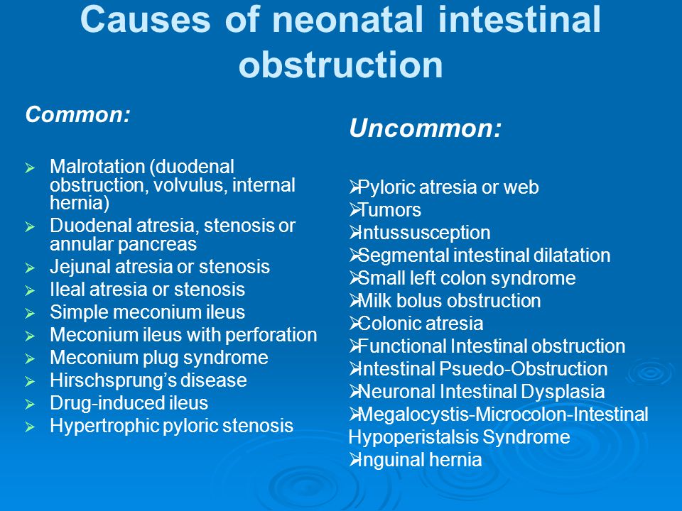
Neonatal intestinal obstruction occurs in 1/1500 live births. Etiologies are from intrinsic developmental defects, abnormalities of peristalsis or abnormal intestinal contents, or from insults in utero after the formation of normal bowel. Failure to recognize neonatal bowel obstruction can result in aspiration of vomit, sepsis, mid-gut infarction or enterocolitis.
Differential Diagnosis for failure to pass meconium:
Disorders of the small intestine
- Duodenal atresia
- Jejunoileal atresia
- Malrotation and volvulus
- Meconium ileus
Disorders of the large intestine
- Meconium plug syndrome
- Anorectal malformation
- Hirschsprung’s disease
- Small left colon syndrome
Other causes
- Narcotics
- Electrolyte abnormalities; hypermagnesemia, hypokalemia, hypercalcemia
- Hypothyroidism
- Sepsis
- Congestive heart failure
Subsequent obstruction with passage of meconium
- Pyloric Stenosis
Meconium plug syndrome: Meconium plug syndrome is the most common form of functional bowel obstruction in the newborn, with an incidence of 1/500.
It is a transient form of distal colon or rectal obstruction caused by inspissated and dehydrated meconium, the etiology for which is unknown.
Diagnosis is made through barium enema revealing the outline of meconium plug. Barium enema can also be therapeutic along with rectal stimulation in inducing passage of the meconium.
Generally, infants with meconium plug syndrome have normal bowel function after passing the meconium plug.
Anorectal malformation: The incidence of anorectal malformation is 1/4000 live births, including anal stenosis, imperforate anus, and fistula.
Malformations are caused by a defect in embryonal development where the urorectal septum, lateral mesoderm and ectodermal structures combine to form the normal rectum and lower urinary tract.
70% of infants with anorectal malformation have associated anomalies. The mnemonic VACTERL is used to describe the combination of vertebral defects, anal atresia, cardiac defects, tracheoesophageal fistula, renal defects and radial limb hypoplasia.
Anal stenosis accounts for 20% of anorectal anomalies and treatment is with dilatation.
The mortality rate for patients with anorectal malformation is directly related to associated anomalies.
Malrotation and volvulus: Malrotation of the midgut is caused by a failure of normal bowel rotation. The mid-gut does not complete its normal 290° counterclockwise rotation during embryologic development, resulting in abnormal placement and fixation of the small bowel. This can cause obstruction and sometimes infarction of the small and large bowel, known as volvulus. Volvulus occurs when the small intestine rotates around the superior mesenteric artery, leading to vascular compromise and possible infarction/necrosis of the small bowel. Volvulus usually occurs within the first week of life.
Radiographs may show a “gasless” abdomen, may show signs of intestinal dilation, or may be completely normal.
Treatment is with the Ladd’s procedure involving counterclockwise reduction of the volvulus, release of adhesive bands (Ladd’s bands) to mobilize the duodenum, and appendectomy. Recurrence of mid-gut volvulus after the Ladd’s procedure can occur in up to 10% of cases.
Meconium ileus: This disorder is differentiated from meconium plug syndrome by location of the stool. In meconium ileus, the thick, tenacious bowel is most commonly found in the ileum but occasionally occurs in the jejunum or proximal colon.
90%-95% of patients with meconium ileus have cystic fibrosis. But, only 15% of cystic fibrosis patients will have meconium ileus as neonates. Associated anomalies such as volvulus, jejunoileal atresia, or bowel perforation occur in over half of infants with meconium ileus.
Duodenal atresia: The etiology of duodenal atresia is from failure to recanalize the lumen after the solid phase of intestinal development. This occurs between the 4th and 5th week of gestation. 40% of the patients with duodenal atresia have Down Syndrome. Diagnosis can be made in utero if polyhydramnios is present. Plain films can reveal the “double bubble sign”, and an upper GI series might be necessary to distinguish between malrotation and duodenal atresia. Treatment is with duodenoduodenostomy.
Pyloric Stenosis- A condition of hypertrophy of the pylorus, with elongation and thickening of tissue that progresses to near-complete obstruction of the gastric outlet. It typically arises towards the end of the neonatal period, with most cases arising at 3-6 weeks, but it can present even in the first few months. Rate of 3 in 1,000 live births, more commonly in males (4-6:1).
The presentation of pyloric stenosis is a three- to six-week-old baby who has previously fed well and passed stools, and suddenly begins to have non-bilious, usually projectile vomiting after feeding and demands to be re-fed soon afterwards. An "olive-like" mass is very often described as being palpated in the right upper quadrant of the abdomen. The diagnosis is confirmed by ultrasound examination of the abdomen, which will display a thickening of the pyloric sphincter where the mass is palpated. Patients should be observed for complications of dehydration and vomiting, such as hypochloremic hypokalemic metabolic alkalosis. Fluid resuscitation and surgery are the standard for treatment
Hirchsprung’s disease: Hirchsprung’s disease, or congenital aganglionic megacolon, is a motor disorder of the colon that causes a functional intestinal obstruction. It occurs in 1/5000 infants with a male to female predominance of 4:1. The pathogenesis of the disease is failure of migration of the neural crest cells that form the colonic ganglion cells. Without parasympathetic innervation, the colon cannot relax or undergo peristalsis, resulting in a functional obstruction.
The aganglionic segment is limited to the rectosigmoid in the majority of patients. 10% have full colonic involvement and in 10% more, there is lack of ganglion cells into the small bowel.
Diagnosis of Hirchsprung’s disease can be made with barium enema, revealing a transition zone between the constricted aganglionic segment and the proximal, normally dilated segment.
Confirmation of the diagnosis is made with rectal suction biopsy, which will demonstrate absent ganglion cells, hypertrophied nerve fibers, and elevated acetylcholinesterase activity.
The treatment of Hirchsprung’s disease is through surgical resection of the aganglionic bowel and approximation of normal, ganglionic tissue near the anus.
The major complications of the disease, even after surgical resection, are bowel obstruction and enterocolitis, though most patients go on to normal or near-normal bowel function.
The major complications of the disease, even after surgical resection, are bowel obstruction and enterocolitis, though most patients go on to normal or near-normal bowel function.
References:
- Failure to pass meconium: diagnosing neonatal intestinal obstruction. Loening-Baucke V. Am Fam Physician. Nov 1999; 60(7): 2043-2050.
- Bilious vomiting in the newborn: rapid diagnosis of intestinal obstruction. Kimura K. Am Fam Physician. May 2000; 61(9): 2791-2798.
- Imaging of neonatal gastrointestinal obstruction. Hernanz-Schulman M. Radiol Clin North Am. Nov 1999; 37(6):1163-1186.
- Behrman: Nelson Textbook of Pediatrics, 16th ed (2002). Philadelphia: W.B. Saunders Company.
- A decade of experience with the primary pull-through for Hirschsprung disease in the newborn period: a multicenter analysis of outcomes. Teitelbaum DH. Ann Surg. Sep 2000; 232(3):372-380.
- Genetics of Hirschsprung's disease. Parisi MA. Curr Opin Pediatr. Dec 2000; 12(6):610-617.
- Long-term outcome and quality of life after the Swenson procedure for Hirschsprung's disease. Bai Y, Chen H, Hao J, et al. J Pediatr Surg 2002; 37:639.
- Ultrasonographic diagnosis criteria using scoring for hypertrophic pyloric stenosis. Ito S, Tamura K, Nagae I, et al. J Pediatr Surg 2000; 35:1714.
- NEJM.A Newborn Boy with Vomiting,Diarrhea, andAbdominal distention. Jan 26, 2012
Did you realize there's a 12 word sentence you can speak to your partner... that will induce deep emotions of love and impulsive attraction for you deep inside his chest?
ReplyDeleteThat's because hidden in these 12 words is a "secret signal" that triggers a man's impulse to love, look after and guard you with his entire heart...
12 Words Who Fuel A Man's Love Instinct
This impulse is so built-in to a man's genetics that it will drive him to work better than before to do his best at looking after your relationship.
Matter-of-fact, fueling this influential impulse is absolutely important to getting the best possible relationship with your man that the instance you send your man a "Secret Signal"...
...You'll soon find him open his soul and heart for you in a way he haven't experienced before and he will recognize you as the only woman in the galaxy who has ever truly attracted him.